(Updated 4/9/2022) – I would like to introduce to you a real life demonic type entity that grows inside of both humans and animals. It can form eyes, teeth, hair and even organs as it parasitically feeds on your body for its life.
In modern science, we call it by the name “teratoma tumor” which is Greek for monster, and the Latin tumor, from tumere ‘to swell’ which refers to a mass that grows somewhere in the body.
As you can see for yourself, these tumors look like the scariest fricken monsters that I have ever seen and they are growing in people.
These tumors contain several types of cells and began to grow various forms of tissue such as hair, teeth, eyeballs and even organs and feet. These things are real-life entities growing within people!
Teratomas are uncommon in children, but they can happen at any age. They can be cancerous (malignant) or not cancerous (benign).
Doctors aren’t sure what causes teratomas, but they may be related to genetic changes.
Teratomas are considered a type of germ cell tumor. Germ cells are a type of cell present already in our bodies that begin as an immature form of egg or sperm (gametes) and then undergo a process known as differentiation to become specialized cells like skin cells, brain cells or muscle cells.
During this process, these germ cells migrate to different parts of the body where they will eventually differentiate.
However, at some point during this migration process, some germ cells get left behind and do not complete their development into specialized cells.
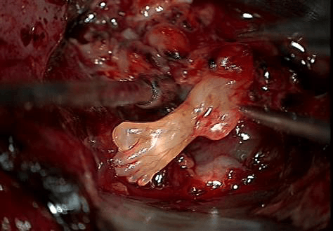
Check out this report in 2008 in Colorado Springs, Colorado of a teratoma case involving a baby with a small tumor in his brain. Doctors operated on a three-day old baby removing the tumor and were shocked to find that it contained one fully-formed foot, another partial foot, and the beginnings of a hand and thigh. The doctor said;
“I’ve never seen anything like it before. It looked like the breach delivery of a baby coming out of the brain.”
There is the case of the 18-year-old Hispanic boy who was overweight and had breathing problems. One day, he had a coughing fit and was rushed to the hospital after his father insisted when he coughed up blood.
The doctors did x-rays revealing what they thought was a massive cyst, the size of a basketball, crushing his lungs. They performed emergency surgery and were shocked when they found a huge teratoma tumor with hair, teeth and what looked like eyes of an underdeveloped fetus.
The dude was literally carrying a monster baby in his body. It is like the invasion of the body snatchers, but in real life.
There are many legends that describe the origin of teratomas. The ancient Greeks, who often attributed anything they did not understand to the gods, thought that these tumors were conceived when a man’s sperm was mixed with menstrual blood.
The modern history of teratoma tumors goes way back to the 16th century.
In the Middle Ages, they called it a “stone baby” and because of that, the condition is sometimes referred to as lithopedion in medical terminology.
It was first reported in 1554 by Italian anatomist Julius Caesar Arantius, who studied a woman with a lump on her neck. He examined it under a microscope and found it contained teeth and hair.
In 1809, French physician Benjamin Bell described ovarian dermoid cysts, which are also known as mature teratomas. It was not until 1827 when Scottish surgeon Robert James Graves coined the term ‘teratoma’ from Greek teras ‘monster’ and onkos ‘tumor’
in the Philippines, people refer to this type of tumor as tikbalang or tikbalang bukol. It’s a Tagalog word referring to a mythical half-horse, half-man creature in Philippine folklore that can cause mischief or death upon encountering humans.
According to the National Cancer Institute, the most common places for these parasitic entities to grow are within the ovaries, testes and the tailbone. The most common form is a dermoid cyst of the ovary, which contains the epidermis, hair follicles, and sebaceous glands.
But sometimes they grow in our heads as in the recent case of this 14-year-old boy with right eye proptosis and visual acuity impairment, headaches, and a neuralgia-like facial pain who was found to have a large tumor filling his right cavernous sinus.
The doctors found that the patient had fat, cartilage, muscle strands, bone, and a primordial tooth.(See link for more info)
Or in the lungs like this modern horror story of the 22-year-old woman with a history of a cough was found to have a mass in the left upper lobe of her lungs, mimicking a fungus ball. Yup, there is that demonic fungus again!
The doctors performed an upper left upper lobectomy revealing a teratoma tumor composed of skin, sebaceous glands, hair follicles, apocrine glands, smooth muscle, cartilage, fat, and respiratory epithelium.(source link)
The good news is that they performed surgery on the young lad and lady to remove the monsters and both lived happily ever after.
There are reports of these tumors all over the internet. Here are some more examples for you to take a deep dive down the demonic tumor rabbit hole.
The New England Journal of Medicine reports that doctors remove the teeth and the tumor from the boy’s head during brain surgery. This is a form of teratoma called fetus in fetu, and may contain highly developed or even parafunctional organs. The NE Journal reports:
“Multiple structures along the right periphery of the mass showed characteristics similar to those of teeth in the mandible (arrows). The patient underwent tumor resection through a right pterional approach; multiple fully formed teeth were seen in the tumor mass (Panel B). On pathological examination, an adamantinomatous craniopharyngioma was identified, which is a slow-growing tumor arising from Rathke’s pouch, an embryonic precursor to the anterior pituitary.
Such tumors consist of organized stratified squamous epithelium and wet keratin (keratin nodules) and may be cystic; the cysts are filled with viscous, yellow fluid containing cholesterol crystals. Histologically, adamantinomatous craniopharyngiomas closely resemble some odontogenic tumors and cysts. In the year since he underwent surgery, the patient has required shunting for bilateral subdural hygromas and receives thyroid and adrenal hormone-replacement therapy. He is making good developmental progress, and as part of his follow-up, he currently undergoes routine MRI.”
Livescience recently reported on one of the oldest cases when Spanish archaeologists found one of these monstor tumors calcified in the pelvis of a 30- year old Roman woman who died 1,600 years ago which had bone and four deformed teeth embedded within it.
In 2007 a Japanese woman had a teratoma tumour containing a lump holding a head, one fully formed eye and a number of internal organs removed from her right ovary.
In February 2014 the New England Journal of Medicine reported that doctors in Maryland in the US found fully-grown teeth growing in a 4-month-old baby boy’s enlarged head.
A 2018 study had shown that Intrapulmonary teratoma was misdiagnosed as aspergilloma is a 52‐year‐old woman who was complaining of intermittent episodes of hemoptysis for over 40 years. Chest computed tomography (CT) showed a cavitary lesion (5.5 × 5.3 cm) in the left upper lobe with heterogeneous components. The radiologic manifestations hinted a fungus ball (Fig (Fig1a).1a). Bronchoscopic biopsy and bronchoalveolar lavage failed to identify tumor cells or pathogens. A serum tuberculosis antibody test indicated no abnormalities.
The initial diagnosis was made by a local physician as Aspergillus infection because of positive results of Aspergillus galactomannan (GM) assay. The patient was administered voriconazole (4 mg/kg, q12h) for two weeks; however, her symptoms failed to alleviate. She was transferred to our hospital and underwent left upper lobectomy via a posterolateral thoracotomy through the fifth intercostal space. The specimen revealed a solid‐cystic mass containing yellow sebaceous material, hair and skin (Fig (Fig1b).1b).
Histopathological examination revealed a mature cystic teratoma lined by squamous epitheliums and sebaceous glands, hair follicles, mature adipose tissue, smooth muscles, cartilage and respiratory epithelium without any malignant cells. A final diagnosis of mature cystic intrapulmonary teratoma was established.
Postoperatively, the patient underwent a smooth recovery and was discharged at postoperative day (Source Link)
The word tumor is a general term for the mass that can be medically benign (generally harmless) or malignant (cancerous) growths which can proliferate the body uncontrollably and then destroy the healthy tissues as they spread systematically in the body.
The word cancer comes to us from the Father of Medicine, Hippocrates who in approximately 400 B.C. was said to have coined the term for these masses (tumors) he discovered growing in patients as karkinos — Greek for the word for crab because cancers “grab on and don’t let go.”
A malignant (cancerous) growth refers to a mass which we call a tumor that has the ability to invade surrounding tissues, metastasize (spread to other organs) and which may eventually lead to the patient’s death if untreated.
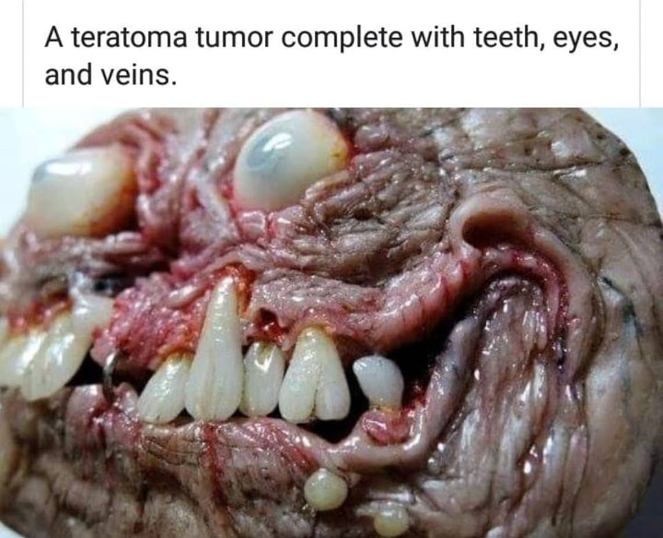
TOP IMAGE SOURCE: Sculpture by Morgan Loebel (@morgansmutations) – This image was used to show what a human looking and fully formed tumor might look like.
Moe is the founder of GnosticWarrior.com. He is a father, husband, author, martial arts black belt, and an expert in Gnosticism, the occult, and esotericism.
It’s exactly why we have survived. These tumors are our body adapting to conditions and failing. Without these mutations we, as a species, would’ve died off before we ever got started.
Fascinating!! I have a dermoid cyst on one of my ovaries. It’s 1-2 cm, and I get it ultrasounded each year. It doesn’t grow. After reading this I want it removed though!!!
Mo, you’ve produced a brilliant information contact with Gnostic Warrior. For me the biggest fight I feel the world has is disseminating the information that Compromise of our political military and national structures are the controlling nature of this earth. If you have any more in depth information regarding the sickly practice of child sacrifice I would be very grateful to hear about it as I feel this is the lynch pin to breaking the demonic controll that surrounds the western world. Interestingly enough child sacrifice does not occur in the East though secret society number are much greater than in the west. For me the three fights I need to war with are
A illegalisation of secret society with criminal code B psychiatric testing to weed out candidates with psychopathic tendencies in positions of government C incarceration for those involved in pedophilic sacrificial child abuse.
If you have advice don’t hesitate to contact. Best wishes to you fine soldier
Very interesting comment- I am so grateful to know there are other people who are aware of the ongoing practices of pedophila, satanic ritual sacrifice, and secret societies that the elite are all members of.
It’s very difficult to know where to begin.
My nephew recently was having a lot of pain in his ear. Turned out, he had a ‘growth’ on his thyroid in his neck that had tentacles that had grown and was entwined around his spine as it pressed against his thyroid gland. His tonsils also had gone bad and had to have them removed also. The growth had a head that appeared to be developing eyes and a mouth and a body with veins that had what appeared to be appendiges. The doctor had no clue what this is, nor did we, until I came across this article. This is the oddest thing I have ever heard of. If not for the fact that he just had surgery to remove this alien looking growth, it would be hard for me to fathom.
It seems as though they may be failed second fetuses…i wonder if any of those cases had history of multiple births in their family. They may have started off as twins but one become grossly under developed for what ever reason. May be some how the embryos failed to detach and one ends up on the inside of the other becoming un able to fully develop forever trapped inside their sibling…not impossible
The desire to get you to read an article that is unremarkable and irrelevant. Or it may be to flush out the conspiracy theorists convinced that it is evidence of devil-worship and human sacrifice. They’re easy to spot, as they are the ones wearing tin foil hats
Just had a thought. In many vaccines there are aborted fetal DNA. They are the WI-38 cell line is a human diploid fibroblast cell line derived from a three month old fetus aborted therapeutically in 1962 in the US. Another cell line, MRC-5, was derived from lung fibroblasts of a 14 week old fetus in 1966 in the United Kingdom. Makes me wonder…if this is some sort of mutation of those cells while inside a living body.
That’s untrue. It says, and I quote: “The word cancer comes to us from the Father of Medicine, Hippocrates who in approximately 400 B.C. was said to have coined the term for these masses (tumors) he discovered growing in patients as karkinos”
I’m a licensed, registered Histopathologist with many years of schooling. This has NOTHING to do with vaccines. How many years of medical school do you have? What licenses do you hold? Are you a member of the American Society for Clinical Pathologists like I am? Have you dissected and studied teratomas before and gotten first hand knowledge or do you just read, adapt and spread false theories from the internet?
CATHERINE NICHOLS POGORZELSKI
on February 15, 2019 at 11:02 pm
No false info here, Giggle fairy, how much time of your supposed eminent educational prowess was actually spent studying vaccines?! Can you name five ingredients?!
And do you actually KNOW how long vaccine ingredients and their adjuvants “spurring them to work,” deposit themselves in the human body and their deleterious effects?! I can name 12 toxic heavy metals that my body has retained I have proof of this or you can check my Facebook page is under Catherine Nichols POGORZELSKI (post fifth chelation) and see just how high that’s just half of it off the chart levels of the following:
gadolinium from MRIs barium from swallow studies, lead, aluminum and mercury from vaccines etc. these are causing my body to hasten to an early grave.
I certainly hope that at least you will ascertain what vaccine ingredients are and make informed consent decisions as to what you ALLOW to be put into your body as a “licensedhealth professional.”
Having already suffered thyroid disease then cancer, bilateral elongated and calcified styloid bone (known as Eagle syndrome-left side removed along with entire thyroid) neurogenic thoracic outlet syndrome, two MTHFR gene mutations, a nerve sheath tumor At C-5/C-6 level, RDD/CRPS, Sjogrens andnits indiced pancreatitis, , Lyme, BABESIA and B.Burgdorferi, Ankylosing Spondylitis, Interstitial cystitis, fibrosis of bowel, carotid stenosis, angiography induced stroke with induced right eye partial blindness, Coccyxdynia, 2 Bowel prolapses and keyhole deformity, bladder prolapse, Vulvodynia, Major depressive disorder, it’s no wonder…
Do you remember the movie with was it Robin Williams playing the father of the child who needed was Lorenzo Eli blues name of it and he got on the web and did research in the early days we don’t need licenses to educate ourselves we go to where the information is that is peer reviewed in the studies and we reduce how do you think I know as a New Jersey housewife to ask for the tests that I had to be able to prove that what I was retaining in my body was what was Making me so sick?! you may need a license to do your job and get paid for it but his patients we don’t need a license to know that what we’re retaining is killing us and we don’t have to allow more bodies were those whom we love to be raped by the toxins injected sometimes from the first day of life, end of discussion !
This is a horrible hypothesis. That is not how vaccines are made. Stop mixing in half truths with your lies. It is true that aborted fetal cells were used to make some of the first vaccines, but only to grow the weakened virus. Its NOT true to imply that they are some how ground up and mixed in with each vaccine. Aborted fetal cells have not been used in more than 50 years. Get your facts straight; there isn’t aborted fetal dna in vaccines.
Human cells from the tissue of aborted fetuses have been used in vaccines since the 1960s, and currently they are used in 11 vaccines. Aborted human fetal cell cultures are used for growing viruses, which are then used in the preparation of inactivated and live virus vaccines. They may also be grown on chick embryos or cells from different animals and bugs, such as army worms.
A study published in 2015 in the journal Human Vaccines & Immunotherapeutics confirms that in addition to the two primary cell cultures that have been used for more than half a century to make hundreds of millions of doses, a third aborted human fetal cell culture may be used. Called Walvax-2, it has been assessed as a culture for growing rabies, hepatitis, and varicella viruses.
Source tissue for this new cell line was obtained from nine fetuses provided by the Department of Obstetrics and Gynecology of Yunnan Hospital in China. Relatively little additional information about Walvax-2 is available from public sources. It is not clear how the name was derived, what government agency or company is leading the research, or the possible application of the cell line to vaccine production.
Read the full article in the NVIC’s online newspaper The Vaccine Reaction. To learn more about how vaccines are created, read Understanding the Rationale Behind Vaccines. Listen to Theresa Deischer, PhD talk about how humans and their parts can be exploited for medical manufacturing and product commodities in The Commoditization of Human Beings.
After getting this link by email, I was of course creep’ed out. I have seen other videos of twin merging but this was possibly different [ or not ]. It brought me back to seeing an exorcist for my “husband” as he became increasing filled with rage, talking to himself, locked in a dark room day and night and continued to grow larger. One evening, watching him come down the stairs, around a corner in which a table sat with a vase on it..no where close to where he was walking but as he came around this corner, the table went down and the vast broke on the floor. I literally walked his every step and I am telling you, [ he ] did not touch any of this with his physical body. It was as if he was “layered” in multiple unseen demons that extended outside of his own body. They knocked this over not him. Not one doctor will do any X RAYS of his gut, nor his brain and I want to know why ? My daughter and I had a “Kodak Moment” with him a few years ago sitting in the dining room. Normally if he saw a camera pointed at him he would toss it out a window. She managed to pick mine up with a flash on and took two photos of him while it just sat their within any emotion as if he did not know it was happening. But as seen in the photo, something shows in his eyes……like the dark side of what is inside of him, I can only share seemed so much like Legion or Nebuchadnezzar . He just consistently gets worse. I have never found help for this situation which would also be very hard to share with any normal person, clergy, or anyone. But I have the photos, and it is very real. I often wonder what is inside of him and that experience with the vase made it clear for me that the raging and talking to [ no one human ] has to be a demonic force that is inside of him. Any thoughts on this are welcome ……we will lose our home to foreclosure on MAY DAY -due to MERS FRAUD because I can not get him to sign anything or help with anything anymore. He is in his own world in that room.
Demonic spirits are real. Keep the faith, pray, fast and speak with someone who has a deliverance ministry. I’ve not encountered demonic spirits in the states as I did while in India on a week long revival but they do exist. Prayers for you and your family.
Hello. I can help. I have demonic possession myself. And you are exactly right the demons knocked down the table and vase. My husband also treats me and a lot of other suffering people.
prevention of this? Never again enter into any agreement to embody in
any universe or planet of pain.
This is the lowest vibratory realm except one of the satellite bodies called
“moons”.
And who feeds on human bodies? Someone from somewhere or everyone
from everywhere. Human life is beyond sad…beyond pathetic. And we are
right in the crosshairs. Thanks God.
It’s not! The first photo is actually a sculpted belt buckle. The artist found this article, screen shot it, and posted it to his Instagram account, laughing about how it was mistaken for a teratoma tumor. That’s how I found it. (He’s @morgansmutations on IG.)
It isn’t. Teratoma tumours do not look like that. That photo is actually of a belt buckle made by artist Morgan Loebel. You can find his work on Instagram.


wtf causes this thing to grow? There’s no way in hell this is normal, otherwise we as a species would be extinct.
WHAT CAUSES THIS?!
It’s exactly why we have survived. These tumors are our body adapting to conditions and failing. Without these mutations we, as a species, would’ve died off before we ever got started.
I just watched in horror , yes I’m behind in TV, about this … I couldn’t believe my eyes!!! This thing has eyes? Teeth? Veins? OMG
Detailed Notes on case
Fascinating!! I have a dermoid cyst on one of my ovaries. It’s 1-2 cm, and I get it ultrasounded each year. It doesn’t grow. After reading this I want it removed though!!!
Joey Ramone had a teratoma on his spine.
I was born with a teratoma tumor in my spine..mine had hair and wax…but caused me a lot of problems that I’m stuck with
My left nut grew hands. It gives me hand jobs every night before bed.
nice
Ha ha
🙄
Mo, you’ve produced a brilliant information contact with Gnostic Warrior. For me the biggest fight I feel the world has is disseminating the information that Compromise of our political military and national structures are the controlling nature of this earth. If you have any more in depth information regarding the sickly practice of child sacrifice I would be very grateful to hear about it as I feel this is the lynch pin to breaking the demonic controll that surrounds the western world. Interestingly enough child sacrifice does not occur in the East though secret society number are much greater than in the west. For me the three fights I need to war with are
A illegalisation of secret society with criminal code B psychiatric testing to weed out candidates with psychopathic tendencies in positions of government C incarceration for those involved in pedophilic sacrificial child abuse.
If you have advice don’t hesitate to contact. Best wishes to you fine soldier
Very interesting comment- I am so grateful to know there are other people who are aware of the ongoing practices of pedophila, satanic ritual sacrifice, and secret societies that the elite are all members of.
It’s very difficult to know where to begin.
What do you mean I’m sorry for my ignorance !!
That first image is actually a belt buckle made by an artist who enjoys “teratoma art”. Cool read though.
Elizabeth Wallace
+ would love to talk…
[email protected]
My nephew recently was having a lot of pain in his ear. Turned out, he had a ‘growth’ on his thyroid in his neck that had tentacles that had grown and was entwined around his spine as it pressed against his thyroid gland. His tonsils also had gone bad and had to have them removed also. The growth had a head that appeared to be developing eyes and a mouth and a body with veins that had what appeared to be appendiges. The doctor had no clue what this is, nor did we, until I came across this article. This is the oddest thing I have ever heard of. If not for the fact that he just had surgery to remove this alien looking growth, it would be hard for me to fathom.
It seems as though they may be failed second fetuses…i wonder if any of those cases had history of multiple births in their family. They may have started off as twins but one become grossly under developed for what ever reason. May be some how the embryos failed to detach and one ends up on the inside of the other becoming un able to fully develop forever trapped inside their sibling…not impossible
That’s exactly why I thought as soon as I started reading the article.
So what makes it “Demonic” again ?
The desire to get you to read an article that is unremarkable and irrelevant. Or it may be to flush out the conspiracy theorists convinced that it is evidence of devil-worship and human sacrifice. They’re easy to spot, as they are the ones wearing tin foil hats
Good question.
It’s ugliness
Random chantz is fucked up stupid
I have something living in me
Peace bro this is , sadness i pray we will leave here soon miss annie
Just had a thought. In many vaccines there are aborted fetal DNA. They are the WI-38 cell line is a human diploid fibroblast cell line derived from a three month old fetus aborted therapeutically in 1962 in the US. Another cell line, MRC-5, was derived from lung fibroblasts of a 14 week old fetus in 1966 in the United Kingdom. Makes me wonder…if this is some sort of mutation of those cells while inside a living body.
Great points!
excellent hypothesis
Rob, if there’s a Greek word for it then they’ve been around for much longer than vaccines. Not to mention this also happens in animals.
Colin, it only states that they’ve taken the name from the Greek word for monster, not that there is a Greek word for this particular phenomena…
That’s untrue. It says, and I quote: “The word cancer comes to us from the Father of Medicine, Hippocrates who in approximately 400 B.C. was said to have coined the term for these masses (tumors) he discovered growing in patients as karkinos”
I’m a licensed, registered Histopathologist with many years of schooling. This has NOTHING to do with vaccines. How many years of medical school do you have? What licenses do you hold? Are you a member of the American Society for Clinical Pathologists like I am? Have you dissected and studied teratomas before and gotten first hand knowledge or do you just read, adapt and spread false theories from the internet?
Odd that a doctor of such caliber would even respond to this and offer still with such vitriol. The hypothesis was reasonable.
Why are u getting so defensive. In actuality it’s make quite a lot of sense. Everybody knows what’s in those disgusting vaccines.
No false info here, Giggle fairy, how much time of your supposed eminent educational prowess was actually spent studying vaccines?! Can you name five ingredients?!
And do you actually KNOW how long vaccine ingredients and their adjuvants “spurring them to work,” deposit themselves in the human body and their deleterious effects?! I can name 12 toxic heavy metals that my body has retained I have proof of this or you can check my Facebook page is under Catherine Nichols POGORZELSKI (post fifth chelation) and see just how high that’s just half of it off the chart levels of the following:
gadolinium from MRIs barium from swallow studies, lead, aluminum and mercury from vaccines etc. these are causing my body to hasten to an early grave.
I certainly hope that at least you will ascertain what vaccine ingredients are and make informed consent decisions as to what you ALLOW to be put into your body as a “licensedhealth professional.”
Having already suffered thyroid disease then cancer, bilateral elongated and calcified styloid bone (known as Eagle syndrome-left side removed along with entire thyroid) neurogenic thoracic outlet syndrome, two MTHFR gene mutations, a nerve sheath tumor At C-5/C-6 level, RDD/CRPS, Sjogrens andnits indiced pancreatitis, , Lyme, BABESIA and B.Burgdorferi, Ankylosing Spondylitis, Interstitial cystitis, fibrosis of bowel, carotid stenosis, angiography induced stroke with induced right eye partial blindness, Coccyxdynia, 2 Bowel prolapses and keyhole deformity, bladder prolapse, Vulvodynia, Major depressive disorder, it’s no wonder…
Do you remember the movie with was it Robin Williams playing the father of the child who needed was Lorenzo Eli blues name of it and he got on the web and did research in the early days we don’t need licenses to educate ourselves we go to where the information is that is peer reviewed in the studies and we reduce how do you think I know as a New Jersey housewife to ask for the tests that I had to be able to prove that what I was retaining in my body was what was Making me so sick?! you may need a license to do your job and get paid for it but his patients we don’t need a license to know that what we’re retaining is killing us and we don’t have to allow more bodies were those whom we love to be raped by the toxins injected sometimes from the first day of life, end of discussion !
Wow. Plausible.
Fantastic! I was thinking stem cells gone awry myself…
oh my god no, not more ridiculous vaccine conspiracy nonsense…
This is a horrible hypothesis. That is not how vaccines are made. Stop mixing in half truths with your lies. It is true that aborted fetal cells were used to make some of the first vaccines, but only to grow the weakened virus. Its NOT true to imply that they are some how ground up and mixed in with each vaccine. Aborted fetal cells have not been used in more than 50 years. Get your facts straight; there isn’t aborted fetal dna in vaccines.
Drew: I believe you are dead wrong about a few things, there being deliberate use of aborted fetal cell lines in vaccines since the 1960’s!
Now that you know this for fact I am sure you will let others know the truth… won’t you?!
https://www.nvic.org/nvic-vaccine-news/january-2018/new-human-fetal-cell-lines-for-vaccine-production.aspx
Human cells from the tissue of aborted fetuses have been used in vaccines since the 1960s, and currently they are used in 11 vaccines. Aborted human fetal cell cultures are used for growing viruses, which are then used in the preparation of inactivated and live virus vaccines. They may also be grown on chick embryos or cells from different animals and bugs, such as army worms.
A study published in 2015 in the journal Human Vaccines & Immunotherapeutics confirms that in addition to the two primary cell cultures that have been used for more than half a century to make hundreds of millions of doses, a third aborted human fetal cell culture may be used. Called Walvax-2, it has been assessed as a culture for growing rabies, hepatitis, and varicella viruses.
Source tissue for this new cell line was obtained from nine fetuses provided by the Department of Obstetrics and Gynecology of Yunnan Hospital in China. Relatively little additional information about Walvax-2 is available from public sources. It is not clear how the name was derived, what government agency or company is leading the research, or the possible application of the cell line to vaccine production.
Read the full article in the NVIC’s online newspaper The Vaccine Reaction. To learn more about how vaccines are created, read Understanding the Rationale Behind Vaccines. Listen to Theresa Deischer, PhD talk about how humans and their parts can be exploited for medical manufacturing and product commodities in The Commoditization of Human Beings.
thank you for correcting the unvaccinated potatos
😂😂😂😂
After getting this link by email, I was of course creep’ed out. I have seen other videos of twin merging but this was possibly different [ or not ]. It brought me back to seeing an exorcist for my “husband” as he became increasing filled with rage, talking to himself, locked in a dark room day and night and continued to grow larger. One evening, watching him come down the stairs, around a corner in which a table sat with a vase on it..no where close to where he was walking but as he came around this corner, the table went down and the vast broke on the floor. I literally walked his every step and I am telling you, [ he ] did not touch any of this with his physical body. It was as if he was “layered” in multiple unseen demons that extended outside of his own body. They knocked this over not him. Not one doctor will do any X RAYS of his gut, nor his brain and I want to know why ? My daughter and I had a “Kodak Moment” with him a few years ago sitting in the dining room. Normally if he saw a camera pointed at him he would toss it out a window. She managed to pick mine up with a flash on and took two photos of him while it just sat their within any emotion as if he did not know it was happening. But as seen in the photo, something shows in his eyes……like the dark side of what is inside of him, I can only share seemed so much like Legion or Nebuchadnezzar . He just consistently gets worse. I have never found help for this situation which would also be very hard to share with any normal person, clergy, or anyone. But I have the photos, and it is very real. I often wonder what is inside of him and that experience with the vase made it clear for me that the raging and talking to [ no one human ] has to be a demonic force that is inside of him. Any thoughts on this are welcome ……we will lose our home to foreclosure on MAY DAY -due to MERS FRAUD because I can not get him to sign anything or help with anything anymore. He is in his own world in that room.
Demonic spirits are real. Keep the faith, pray, fast and speak with someone who has a deliverance ministry. I’ve not encountered demonic spirits in the states as I did while in India on a week long revival but they do exist. Prayers for you and your family.
Why are you all still there?
I will keep you all in prayer.
Can I add you on Facebook?
Demons are real. Get an exorcist/priest ASAP.
Priest, exorcist, prayer…….Is this not a agnostic site??? Should not be a place for prayers nor priests!!
No, it is a Gnostic site which is the opposite of Agnostic.
Contact Russ Dizdar @ the black awakening .com
Hello. I can help. I have demonic possession myself. And you are exactly right the demons knocked down the table and vase. My husband also treats me and a lot of other suffering people.
prevention of this? Never again enter into any agreement to embody in
any universe or planet of pain.
This is the lowest vibratory realm except one of the satellite bodies called
“moons”.
And who feeds on human bodies? Someone from somewhere or everyone
from everywhere. Human life is beyond sad…beyond pathetic. And we are
right in the crosshairs. Thanks God.
Are you sure the topmost photo is an actual picture of a teratoma tumor? Whats with the metal lip piercing?
It’s not! The first photo is actually a sculpted belt buckle. The artist found this article, screen shot it, and posted it to his Instagram account, laughing about how it was mistaken for a teratoma tumor. That’s how I found it. (He’s @morgansmutations on IG.)
It isn’t. Teratoma tumours do not look like that. That photo is actually of a belt buckle made by artist Morgan Loebel. You can find his work on Instagram.
Hahaha what the fuck have you been smoking? Pass it along
Whoa you seem ro have a whole different perspective on this
What did it mean by this