There is a common belief that the calcification or the fluoridation of the pineal gland causes humans to lose their connection to the spiritual world, and they become unenlightened as a result of this mineralization.
In the course of my research into the modern science of our brains, I have found that is simply not true. When the hippocampus or ammon’s horn becomes infested with worms, it begins to mineralize and we lose our connection with past memories, intuition, and enlightenment. This results in the loss of the connection to our third eye and Alzheimer’s Disease that then causes the severe depression of the soul which I’ll explain in more detail below.
After all, this is the part of our brains that modern scientists have determined without a doubt is “critical to learning and remembering relationships that characterize spatial layouts, items in the particular context in which they have been experienced, and other associative, sequential or logical relationships among experiences.”(1) Scientists have also found that the hippocampus is not just filing away memories, it is also a type of internal intuition brain processor that helps us connect with other related memories to give them meaning.
In other words, our hippocampus is simply what we can call our third eye which is the true place of gnosis, and when it becomes damaged, we began to lose one of the most important parts of our brain. This loss of our human memory processor, and soul GPS intuition unit causes us to become third eye blind. A process that sometimes leads to what the modern medical establishment has called, Alzheimer’s disease.
Let me please add that the calcification and fluoridation of the pineal gland can happen just like it does in almost all parts of the brain and body. But it does not cause humans to lose memory, lose their spirituality, gnosis and/or make them unenlightened. In fact, there is no modern scientific evidence and/or studies that can prove that the mineralization of this brain gland causes this.
As I mentioned above, there are a lot of studies and evidence that point to the hippocampus as our third eye, and when it is damaged, we lose our memories. In this article, I would like to share with you some of these studies and research papers so you can explore them yourself. There are also many other articles I have written on this subject that you can click on below this article.
“Our study shows that the prevalence of intrahippocampal calcification at CT increases with age, and intrahippocampal calcification is a relatively common finding in patients older than 50 years, even in patients without evidence of neurodegeneration. This is in contrast to previous radiology literature, which suggests that intrahippocmpal calcification only occurs in rare conditions, such as lipoid proteinosis (3–5) and Taenia solium cysticercosis (6). Knowledge of the agerelated prevalence of intrahippocampal calcification may prevent radiologists from proposing unnecessary differential diagnostic considerations and further investigations, particularly in patients
older than 50 years.”
ATROPHY OF THE HIPPOCAMPUS CAUSES ALZHEIMER’S DISEASE (THIRD EYE SHRINKAGE)
In the early stages of Alzheimer’s Disease (AD), short-term memory begins to fade, and is one of the most frequent causes of dementia, representing 50 to 60% of all dementia cases and affecting 10 to 20% of people older than 65 years (Talmelli et al. 2010). Over approximately the last 10 years, scientists have observed in Alzheimer patients that their hippocampus begins to atrophy as it shrinks in size.
This shrinkage can be compared to when a person who doesn’t use their muscles in their body, and as they get older, they literally began to shrink in size as all their muscles shrink so all that is left is blood, skin and bone from this atrophy. When a person cannot form new memories, like the muscles on their body, their brains muscle known as the hippocampal region begins to die, and so does the patient with this disease.
A study done in 2011 concluded, “Alzheimer disease-specific cortical thinning and hippocampal volume loss are consistent with a sigmoidal pattern, with an acceleration phase during the early stages of the disease.”
In 2009, the medical journal of the American Academy of Neurology had reported on a study by Wouter Henneman, MD, of VU University Medical Center in Amsterdam, The Netherlands that involved 64 people with Alzheimer’s disease. MRI scans were performed on all of the participants. The researchers measured the volume of the whole brain and the hippocampus area, at the beginning and end of the study, and calculated the rate of shrinkage in the brain over that time.
They had found that the people who did not have dementia at the beginning of the study, those with smaller hippocampal volumes and higher rates of shrinkage were two to four times as likely to develop dementia as those with larger volumes and a slower rate of atrophy.
The study’s author, Wouter Henneman, had said about the research, “This finding seems to reflect that at the stage of mild cognitive impairment, considerable atrophy has already occurred in the hippocampus. In people who already have Alzheimer’s disease, the loss of nerve cells is more widespread throughout the brain.”
WORMS TAKE OVER THE HIPPOCAMPUS (THIRD EYE BECOMES THE WORMS EYE)
An interesting note on this study above is that the Taenia solium cysticercosis causes intrahippocampal calcification. Taenia solium cysticercosis is a parasitic tissue infection caused by larval cysts of the tapeworm. The CDC says that these worm larval cysts infect brain, muscle, or other tissue, and are a major cause of adult onset seizures. In the United States, cysticercosis is considered one of the Neglected Parasitic Infections (NPIs), a group of five parasitic diseases that have been targeted by CDC for public health action.
Here is an x-ray showing what these worms do to the brain. It appears to be no different from a worm-eaten fruit, plant, tree or rotting animal carcass.
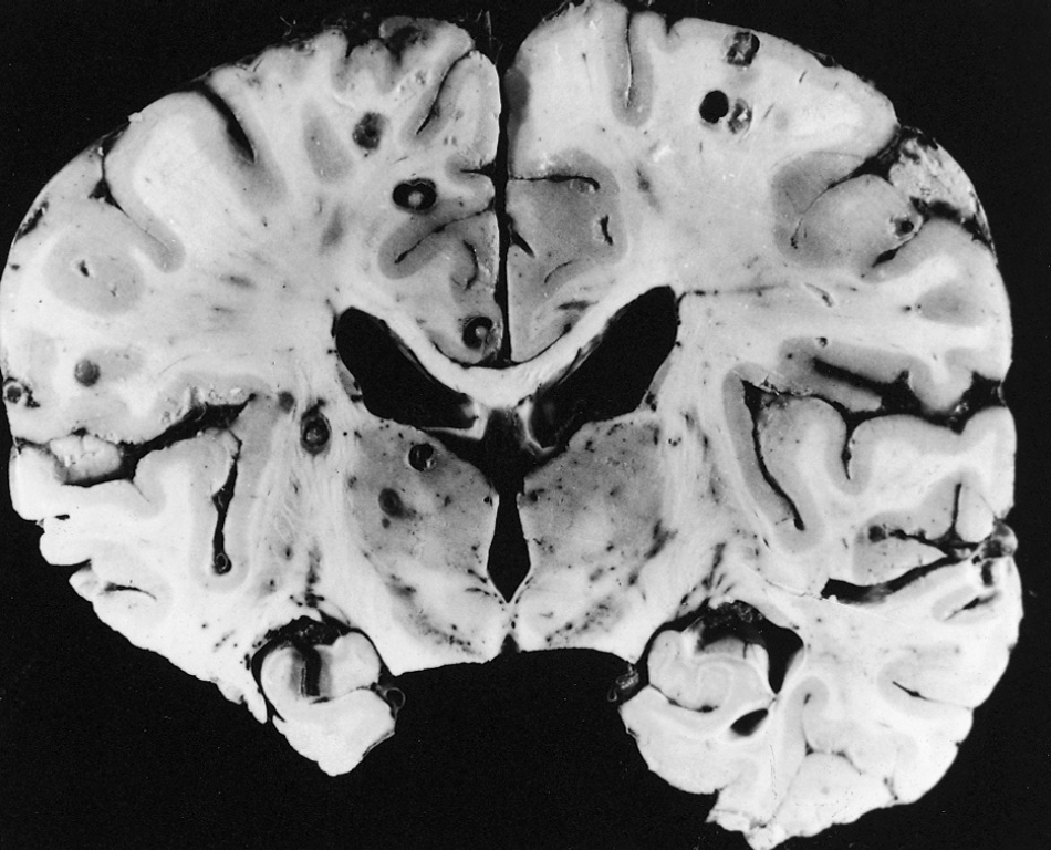
Here is an image of Neurocysticercosis, vesicular stage, in a Mexican adult.
What I believe is that many people who lose their memories and minds are being eaten out of their temples (skulls) by what the church had labeled demons, and their king, the devil who Dante had called a worm. These devilish blood born demon worms who eat our bodies and brains I have written about many times before here on the Gnostic Warrior.
Perhaps these worms are the main cause of Alzheimer’s Disease and in fact, all disease…
Moe is the founder of GnosticWarrior.com. He is a father, husband, author, martial arts black belt, and an expert in Gnosticism, the occult, and esotericism.
 humans to lose their connection to the spiritual world, and they become unenlightened as a result of this mineralization.
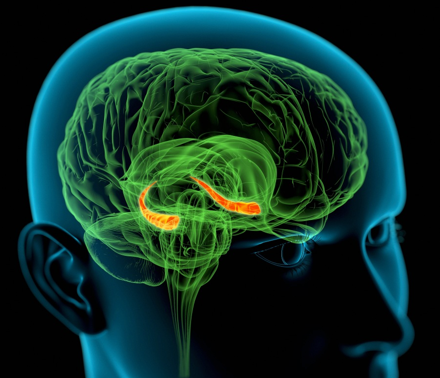 After all, this is the part of our brains that modern scientists have determined without a doubt is “critical to learning and remembering relationships that characterize spatial layouts, items in the particular context in which they have been experienced, and other associative, sequential or logical relationships among experiences.”(1) Scientists have also found that the hippocampus is not just filing away memories, it is also a type of internal intuition brain processor that helps us connect with other related memories to give them meaning.
 SHRINKAGE)
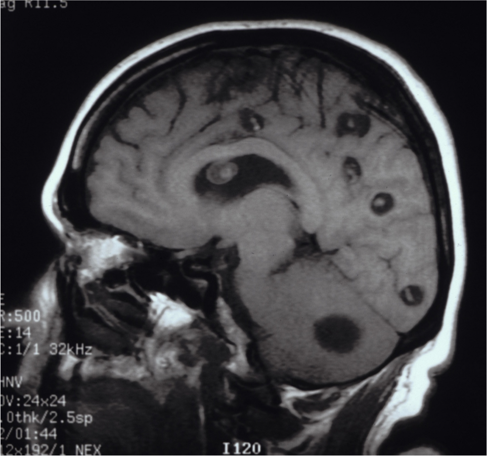


Carl Zimmer – Parasite Rex
Another great article. Curious as to what can be done to eliminate these parasites?
Turpentine